
Back pain and Altered Mental Status
Presentation
A 45-year-old male, with past medical history insulin-dependent diabetes, hypertension, hyperlipidemia, scoliosis was BIBA to the emergency department with altered mental status and respiratory failure.
Per the patient’s wife, over the past year, he has had bouts of occasional chest pain. Seven days prior to arrival he presented to ED with worsening lower back pain, and was discharged with a prescription for steroids/muscle relaxants after negative back X-ray imaging. At that time, patient did not have bowel/bladder incontinence and did not have urine retention; he was able to ambulate. Four days prior to repeat presentation, he became increasingly weak and off balance, spending most the day in bed and was only able to ambulate with assistance. Two days prior to repeat presentation, he became increasingly altered, and started saying "weird things," including odd word substitutions. His wife denied vision/hearing difficulty and facial droop. That evening, he became "hot" and diaphoretic, and was given tylenol without significant improvement. Wife states that she attempted to get him to come to the hospital, but he did not want to do so.
On the day of presentation, the patient became increasingly altered, which led to wife calling 911. EMR reports that they found patient febrile with a temperature of 102, with an O2 sat of 93 to 94% on 4 L O2. Patient decompensated en route and upon arrival to the ED, patient is requiring BVM ventilation by EMS.
Further information obtained from wife revealed that the patient does have an active tooth infection which he has tolerated for the past month, which he has not received dental care for due to cost/insurance difficulties. She endorses patient's cigarette use of 1 pack/day and marijuana use, but denies additional recreational drugs, including no IV drug use.
Initial Vitals
T 39.2 C HR 133 BP 114/70 RR 26 O2 sat 80% on BVM.
Physical Exam
GEN: Patient does open his eyes when his name is called. He does not verbalize. He has shallow respirations with an intact gag reflex. Moving all extremities..
HEENT
-Head: NC/AT;
-Eyes: PERRL, EOMI. No discharge or redness;
-Ears: External ears are normal.
-Nose: Normal nares.
-Mouth and throat: ETT secured. Poor dentition.
NECK: no masses
CV: 4/6 systolic murmuc, regular, tachycardic .
LUNGS: CTAB, no w/r/c
ABD: NBS, no masses or organomegaly.
SKIN: clammy, diaphoretic. No skin rashes or abnormal lesions.
MSK: No deformities.
EXT: No clubbing, cyanosis, or edema.
NEURO: Patient is awake but difficult to arouse, nonverbal, moviang all extremities, sensation appears to be grossly intact, speech is nonverbal, gait examination is deferred.
Workup/ED Course
Rapid sequence intubation was performed, and patient was placed on sedation. Initial ABG showed pH 7.12, Co2 72, pO2 123, Bicarb 22.9. Labs are significant for leukocytosis of 17.7, hyponatremia of 120, hypochloremia of 86, BUN of 30, creatinine of 1.9, glucose of 356. On arrival, code stroke was activated given patient's AMS, but all code stroke CTs were negative.
Sepsis criteria was met with lactate of 3.2, and patient was started on Unasyn and Gentamicin. Patient EKG showed sinus tachycardia, and elevated BNP of 4839, and a troponin of 1517. CT abdomen pelvis showed splenic and renal infarcts likely from thromboembolic phenomenon.
Bedside echocardiogram was performed, showing the following:
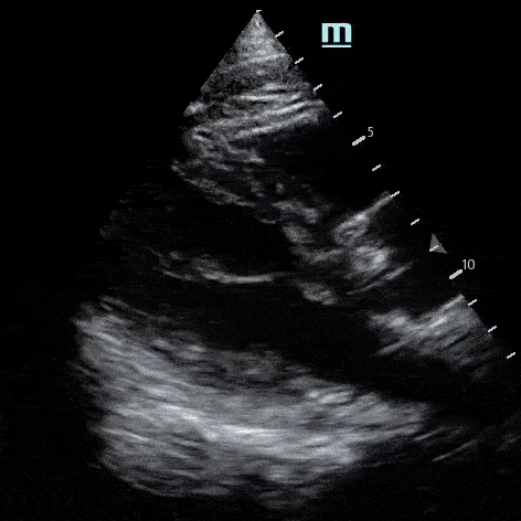
Bedside echo: “Hyperechoic structure seen on aortic valve possible vegetation vs calcification”
Diagnosis/Hospital Course:
The patient was admitted to the ICU on broad spectrum antibiotics for aortic valve endocarditis, which was later confirmed by comprehensive echocardiography, with an additional finding of a bicuspid aortic valve.
MRI of the lumbar spine revealed an epidural abscess significantly narrowing the central canal and compressing the lower cord. Neurosurgery was consulted, and patient underwent abscess drainage. Cardiothoracic surgery was also consulted for aortic valve endocarditis and recommended medical treatment. The patient was eventually extubated and downgraded from the ICU. The patient was also found to have multiple thromboembolic strokes to the frontal and parietal lobes secondary to septic emboli originating from aortic valve endocarditis. The patient received extensive physical and occupational therapy and the time of discharge to acute rehab had regained full 5/5 strength and intact sensation in all extremities and was awake and oriented, speaking in short sentences.
Outcome/Discussion
This patient’s imaging findings are consistent with Aortic Valve Endocarditis with septic emboli, causing multiple thromboembolic strokes and epidural abscess with cord compression.
Takeaway Points
Know risk factors for endocarditis: this patient’s included bicuspid aortic valve and active periodontal disease
Provide return precautions for back pain discharges, including fever, incontinence, or altered mental status
Socioeconomic determinants of health include lack of dental access/coverage, which may have contributed to this patient’s case
Performing early bedside ultrasound can help quickly identify diagnoses and improve care!
Have a cool case that you would like to share? Please email thomas.rauser@uhsinc.com or scan the QR codes in the conference room or TVH ED. Cases will be written with provider anonymity unless consent is given otherwise.